AP Biology Unit 4 Review: Cell Communication and Cell Cycle
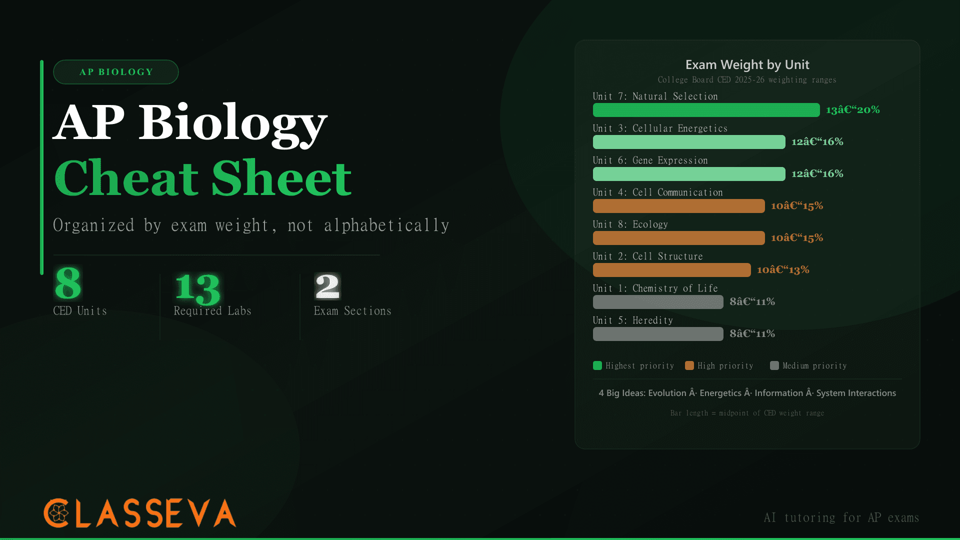
Of all eight units in the AP Biology CED, Unit 4 draws one of the strongest connections between molecular mechanisms and real-world consequences. Signal transduction drives everything from insulin response to cancer initiation; the cell cycle determines whether a cell divides correctly or spirals into uncontrolled growth. At 10–15% of the AP Biology exam, Unit 4 carries more weight than Unit 1 (Chemistry of Life, 8–11%) and matches Unit 8 (Ecology) in exam importance.
Tracing through the last five years of released AP Biology FRQs, what stands out about Unit 4 is how consistently the exam rewards process over vocabulary. The question rarely asks you to define signal transduction. It asks you to trace a specific pathway from ligand binding to cellular response, predict what happens when a receptor is blocked, or explain why a mutation in a checkpoint protein raises cancer risk. The difference between a 3 and a 5 on Unit 4 content is almost always the ability to write a step-by-step causal sequence, not just recall a list of terms. As explained in the AP Biology difficulty breakdown, Cell Communication ranks among the units that most separate high scorers from the field.
This AP Biology Unit 4 review covers both topic clusters in full, with four animated diagrams that trace the pathway and cell cycle mechanisms visually, a complete worked example of the cAMP-PKA signaling cascade, and a full cell cycle phase table mapped to checkpoints.
What's Covered in AP Biology Unit 4?
Unit 4 (Cell Communication and Cell Cycle) covers two interconnected topic clusters and accounts for 10–15% of your AP Biology exam score. The College Board's 2025 AP Biology CED assigns Unit 4 roughly the same weight as Unit 8 (Ecology) and more weight than Unit 1 (Chemistry of Life, 8–11%). AP Biology Unit 4 review material deserves more study time than most students give it.
The Two Topic Clusters
Signal transduction covers how cells receive external signals, relay them through molecular cascades, and produce appropriate responses. The cell cycle covers how cells grow, replicate their DNA, divide via mitosis, and regulate those processes through checkpoints.
These two clusters connect at a specific seam: cell cycle progression depends on signal transduction. Growth factors bind surface receptors, trigger intracellular cascades, and ultimately drive cells through the G1/S checkpoint into S phase. Disruptions to that connection explain how many cancers originate.
How Do Cells Communicate With Each Other?
Cells communicate by releasing chemical signals called ligands, which bind to specific receptor proteins on or inside target cells. The receptor type determines the signaling mechanism: surface receptors handle water-soluble ligands (like peptide hormones and neurotransmitters) that cannot cross the plasma membrane; intracellular receptors handle lipid-soluble ligands (like steroid hormones) that pass through the membrane directly.
Receptor Types and Ligand Binding
The 2025 AP Biology CED focuses on three classes of surface receptor: G-protein-coupled receptors (GPCRs), receptor tyrosine kinases (RTKs), and ligand-gated ion channels. GPCRs are the most tested because they connect to amplification cascades involving second messengers. RTKs dimerize upon ligand binding and phosphorylate each other, triggering growth-related cascades.
Binding specificity matters because it produces tissue-specific responses. Epinephrine triggers glycogen breakdown in liver cells and cardiac muscle contraction in heart cells, even though the epinephrine molecule is chemically identical. The difference lies in which downstream proteins each cell type expresses after the receptor is activated.
Feedback Mechanisms
Negative feedback loops maintain homeostasis by reducing signal output once the response reaches a threshold. The clearest AP exam example is insulin signaling: rising blood glucose triggers insulin release from pancreatic beta cells, insulin drives glucose uptake into liver and muscle cells, blood glucose falls, and reduced blood glucose shuts off further insulin secretion. The loop closes on itself.
Positive feedback loops amplify output toward a discrete endpoint. Childbirth contractions provide the standard biology example, but AP Biology FRQs have also tested positive feedback in the context of caspase activation during apoptosis, where each activated caspase cleaves and activates more caspases.
How to Trace a Signal Transduction Pathway on an FRQ
Every signal transduction pathway, regardless of the specific ligand or receptor, follows three sequential steps: reception, transduction, and response. AP FRQ graders award points for tracing each step in causal order. Knowing the framework as a sequential process matters more than memorizing which proteins belong to which pathway, because the exam regularly presents unfamiliar pathways and asks you to apply the same logic.
The Three-Step Framework
Reception
A ligand (signal molecule) binds to its specific receptor, changing the receptor's shape. For surface receptors like GPCRs, this conformational change activates the receptor's intracellular domain without the ligand entering the cell.
Transduction
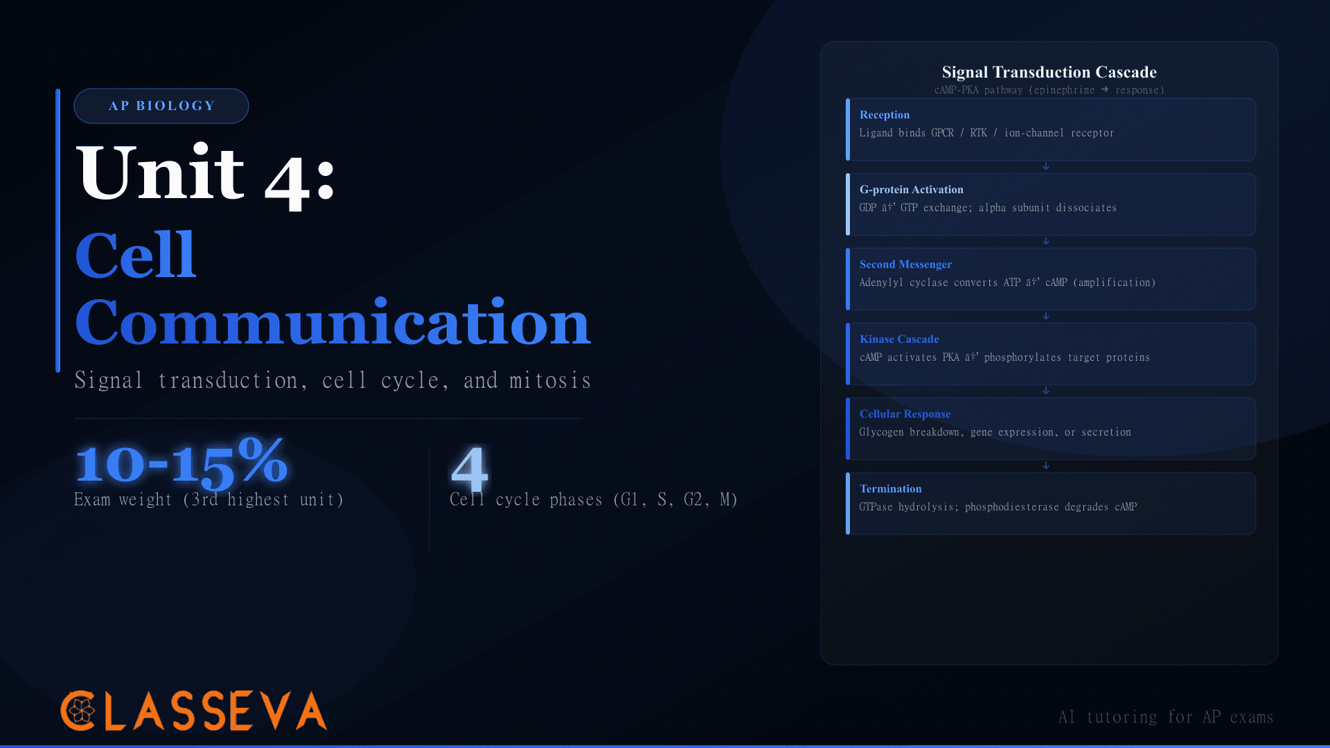
The activated receptor triggers a cascade of molecular events. In GPCR pathways, the receptor activates a G-protein, which activates an effector enzyme like adenylyl cyclase, which produces a second messenger like cAMP. Second messengers amplify the signal by activating many protein kinases simultaneously.
Response
The final activated molecule produces the cellular response: a gene is transcribed, an enzyme is activated or inhibited, or the cell changes shape, secretes a product, or divides.
Worked Example: Epinephrine and the cAMP Cascade
Epinephrine provides the standard AP Biology signal transduction example. An epinephrine molecule binds to a GPCR on the liver cell surface. Binding causes the receptor to activate a G-protein: the alpha subunit detaches and carries GTP. The activated G-protein diffuses along the inner membrane surface until it contacts and activates adenylyl cyclase.
Adenylyl cyclase converts ATP to cyclic AMP (cAMP), the second messenger. One activated adenylyl cyclase molecule produces many cAMP molecules — the amplification step that gives cell signaling its speed and power. Each cAMP molecule activates protein kinase A (PKA), which phosphorylates glycogen phosphorylase, activating it. Glycogen phosphorylase catalyzes glycogen breakdown, releasing glucose into the bloodstream in response to the original stress signal.
When asked to “describe the mechanism by which” a signal produces a response, write a step-by-step causal sequence: “X binds to Y, which causes Z to activate, resulting in...” Never list steps without connecting them causally. FRQ graders look for the word “activates,” “phosphorylates,” or “converts” between each step, not just a list of molecule names.
What Are the Cell Cycle Phases?
The cell cycle has two major stages: interphase and M phase. Interphase contains three sub-phases (G1, S, G2); M phase contains mitosis and cytokinesis. A typical mammalian cell spends roughly 90% of its cycle in interphase and about 10% in M phase — which means any given cell you examine in a tissue slide is almost certainly in interphase, not dividing.
Interphase vs M Phase
| Phase | What Happens | Checkpoint? |
|---|---|---|
| G1 (Gap 1) | Cell grows, synthesizes proteins, monitors nutrient availability | G1/S restriction point — most important |
| S (Synthesis) | DNA replication: genome copied, sister chromatids joined at centromere | S-phase checkpoint monitors replication errors |
| G2 (Gap 2) | Further growth, synthesis of mitotic proteins (cyclins, tubulin) | G2/M checkpoint verifies DNA replication complete |
| Mitosis | Nuclear division into two identical nuclei (5 stages) | Spindle assembly checkpoint (mid-metaphase) |
| Cytokinesis | Cytoplasm divides, producing two daughter cells | None — cell is already committed |
Source: College Board AP Biology CED 2025–26, Unit 4 learning objectives
How Does Mitosis Work? (And How to Identify Each Phase)
Mitosis divides one nucleus into two genetically identical nuclei through five stages: prophase, prometaphase, metaphase, anaphase, and telophase. AP exam questions routinely show a cell image or description and ask students to name the stage from chromosome position or spindle arrangement. Knowing the visual signature of each phase — not just the definition — is the testable skill here.
The Stages of Mitosis
Chromosomes aligned at center = Metaphase. Chromosomes moving to poles = Anaphase. Nuclear envelope visible around condensed chromosomes = Telophase. No visible chromosomes (diffuse chromatin) = Interphase. Spindle attached but chromosomes still scattered = Prometaphase.
How Do Cell Cycle Checkpoints Prevent Cancer?
Three checkpoints verify that the cell meets specific requirements before proceeding to the next phase. All three operate through the same mechanism: specific proteins hold progression blocked until conditions are met, at which point checkpoint proteins are inactivated and the cycle continues.
Cyclins and CDKs
Cyclin-dependent kinases (CDKs) are the enzymes that drive phase transitions, but they only become active when bound to partner cyclin proteins. CDK proteins stay at constant levels throughout the cycle; cyclin concentrations rise and fall, creating timed windows of CDK activity that open and close checkpoints.
The most tested example in AP Biology FRQs is cyclin B combined with CDK1 to form MPF (M-phase promoting factor). MPF drives the G2-to-M transition by phosphorylating nuclear envelope proteins (which dissolve the envelope) and condensin proteins (which condense chromatin). As MPF triggers mitotic entry, it activates enzymes that degrade cyclin B, removing MPF activity and ensuring the cell moves through M phase rather than staying stuck in it.
When Checkpoints Fail
The G1/S checkpoint, also called the restriction point, is the point of commitment. A cell that passes it will complete division regardless of later conditions. The retinoblastoma protein (Rb) holds this checkpoint closed by binding a transcription factor needed for S-phase genes. When CDK activity phosphorylates Rb, the transcription factor is released, and S-phase gene expression begins.
The protein p53, encoded by TP53, monitors DNA damage at both the G1/S and G2/M checkpoints. When p53 detects damage, it activates the gene for p21, a CDK inhibitor that halts the cycle. Cells with intact p53 either repair damage before proceeding or trigger apoptosis. Cells with mutant TP53 cannot stop cycling after DNA damage, accumulate additional mutations with each division, and may develop into tumors.
Apoptosis: When Cells Die on Purpose
Apoptosis is programmed cell death — a controlled dismantling of the cell that prevents the tissue damage caused by uncontrolled cell rupture (necrosis). The process requires energy and proceeds through a caspase protease cascade. Once initiated, caspase activation self-amplifies and becomes irreversible.
AP Biology places apoptosis in Unit 4 because it functions as a cell cycle outcome: when checkpoints detect irreparable damage, a cell can either arrest permanently (senescence) or enter apoptosis. The Bcl-2 family of proteins regulates whether the mitochondria release cytochrome c, which initiates the caspase cascade. Many cancers overexpress Bcl-2, suppressing apoptosis and allowing damaged cells to survive and divide indefinitely. Several classes of cancer drugs target Bcl-2 to restore apoptotic sensitivity.
Apoptosis: controlled, requires ATP, cell shrinks, organelles stay intact, phagocytes clean up the fragments. Necrosis: uncontrolled, cell swells and ruptures, contents leak into surrounding tissue, triggers inflammation. AP FRQs distinguish these by asking whether a cell death scenario is “programmed” or “pathological”.
How Is Unit 4 Tested on the AP Exam?
Unit 4 appears on the AP Biology exam in both sections. Signal transduction pathway tracing has appeared in some form on four of the last five released AP Biology FRQs. The 2025 FRQ Question 2 tested signal transduction using moth pheromone receptor activation, confirming that the exam applies the same three-step framework to unfamiliar biological contexts. Knowing the framework matters more than memorizing any single pathway name.
Cell cycle questions appear most often as MCQs asking students to identify a phase from a description or graph, or to predict the result of a checkpoint protein mutation. Short FRQs have tested what happens when cyclin B is overexpressed (cells skip G2/M checkpoint and enter M phase prematurely) or when p53 is lost (cells fail to arrest after DNA damage and accumulate mutations).
Mitosis vs Meiosis: Preview of Unit 5
Mitosis
- •Purpose: growth and tissue repair
- •Produces 2 genetically identical cells
- •One division (prophase → cytokinesis)
- •Occurs in somatic (body) cells
- •Daughter cells are diploid (2n)
Meiosis
- •Purpose: sexual reproduction
- •Produces 4 genetically unique cells
- •Two divisions (Meiosis I + II)
- •Occurs in germ cells (gonads)
- •Daughter cells are haploid (n)
The AP Biology cheat sheet covers both Unit 4 and all seven other units mapped by exam weight. For practice problems on signal transduction pathway tracing and cell cycle identification, the AP Biology practice questions collection includes full FRQ-style worked examples with grading rubrics.
If you're estimating your AP Biology score before exam day, try the tool below. It uses your practice performance to project a score range and flags which units need the most work before May.
AP Score Predictor
Enter your practice test results to estimate your AP Biology score and identify which units — including Unit 4 — need the most attention before exam day.
Key Takeaways
- AP Biology Unit 4 (Cell Communication and Cell Cycle) accounts for 10–15% of the exam, matching Unit 8 (Ecology) as one of the most heavily weighted units outside the top two.
- Every signal transduction pathway follows three steps in sequence: reception (ligand binds receptor), transduction (intracellular cascade with amplification), and response (cellular output). FRQ graders score step-by-step causal language, not vocabulary lists.
- The cAMP-PKA pathway (epinephrine signaling) is the highest-yield worked example: GPCR activation → G-protein → adenylyl cyclase → cAMP (second messenger, amplified) → PKA → response.
- The cell cycle runs through G1, S, G2, and M phase (mitosis + cytokinesis). Mammalian cells spend roughly 90% of their time in interphase. DNA replication occurs only in S phase.
- Three checkpoints control cell cycle progression. The G1/S restriction point (controlled by Rb and CDKs) is the most consequential because passing it commits the cell to division.
- p53 monitors DNA damage at both G1/S and G2/M checkpoints. Loss of TP53 function occurs in over 50% of human cancers. Cyclin B/CDK1 (MPF) drives the G2-to-M transition and degrades rapidly to ensure the cell exits M phase.
- Apoptosis is programmed cell death via caspase cascades. Bcl-2 family proteins regulate whether apoptosis is triggered. Cancer often involves mutations that suppress apoptosis, allowing checkpoint-defective cells to survive.